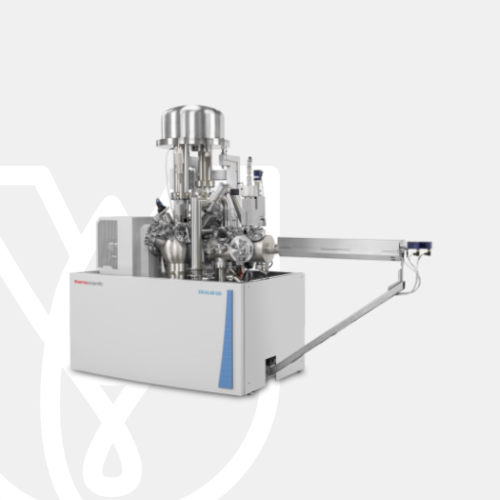
PT Wadya Prima Mulia as the Authorized Distributor for ThermoFisher Scientific in Indonesia, provides ESCALAB QXi X-ray Photoelectron Spectrometer Microprobe
X-Ray phMulti-technique surface analysis instrument with high-resolution X-ray photoelectron spectroscopy and imaging.
ESCALAB QXi X-ray Photoelectron Spectrometer Microprobe
Meet your demands for increased analytical performance and flexibility with the Thermo Scientific™ ESCALAB™ QXi X-ray Photoelectron Spectrometer (XPS) Microprobe, which combines high spectral sensitivity and resolution with quantitative imaging and multi-technique capabilities.
The ESCALAB QXi XPS Microprobe is an expandable and optimized multi-technique instrument with unparalleled flexibility and configurability. It is extremely sensitive, producing high-quality spectra in seconds. System control, data acquisition, processing, and reporting are seamlessly integrated in the powerful Thermo Scientific Avantage Data System. The cutting-edge technology, driven by intuitive software, guarantees world-class results and productivity. The ESCALAB QXi XPS Microprobe, with its unique dual detector system, also delivers superb XPS imaging with excellent spatial resolution.
Monochromated XPS
The twin-crystal, micro-focusing monochromator has a 500 mm Rowland circle, uses an Al anode (or a dual Al and Ag anode with the dual monochromator option), and allows you to select any sample spot size ranging from 200 µm to 900 µm.

Flood electron source
An electron source, co-axial with the analyzer input lens, is used for charge compensation when analyzing non-conducting samples with the monochromatic X-ray source, while a second flood source produces both low-energy ions to assist in providing effective charge compensation and low-energy electrons when the magnetic lens is not in use.

Lens and analyzer
The lens and analyzer system on the ESCALAB QXi XPS Microprobe is optimized for both spectroscopy and for parallel imaging; the single analyzer path means that the same instrument parameters (e.g., pass energy) can be used for both spectroscopy and imaging.

Detectors
The ESCALAB QXi XPS Microprobe is fitted with two detector systems: one optimized for spectroscopy, consisting of an array of six-channel electron multipliers, and one for parallel imaging, consisting of a pair of channel plates and a continuous position-sensitive detector.

Ion gun
The ESCALAB QXi XPS Microprobe has two options for rapid, high-resolution depth profiling: the standard EX06 ion gun, which is optimized for monatomic ion sputtering and ion scattering spectroscopy; and the optional monatomic and gas cluster ion source, MAGCIS, which is capable of monatomic ion profiling, cluster ion profiling, and ion scattering spectroscopy.

Digital Control
All analytical functions are controlled from the Windows Software-based Avantage data system, meaning that the entire analysis process can be performed remotely, if required.

Alignment and calibration
A standards block, which has samples of copper, silver, and gold, can be used for assessing sensitivity, setting the linearity of the analyzer energy scale, calibrating the ion source, aligning the X-ray monochromator, and determining the transmission function of the analyzer.

Sample alignment
All axes of movement on the sample stage are controlled by the Avantage Data System, and a high-resolution digital video camera is fitted to the instrument and is accurately aligned with the analysis position.

Sample manipulator
The computer-controlled, 5-axis, high-precision translator (HPT) allows accurate sample alignment for analysis. When coupled with the new automated sample loading system, it can be used to automatically exchange sample holders and run queued experiments.

Vacuum system
The analysis chamber is constructed from 5 mm-thick mu-metal to maximize the efficiency of the magnetic shielding, and the chamber is pumped using both a turbomolecular pump and a titanium sublimation pump, allowing the analysis chamber to achieve a vacuum better than 5 x 10⁻¹⁰ mbar.

Preparation chambers
The standard Preploc chamber, which is a combined sample entry lock and preparation chamber, has ports that accommodate a variety of sample preparation devices, such as heating/cooling probes, ion guns, high-pressure gas cells, sample parking, and gas admission.

Connectivity
Measurement coordinates can be imported into Avantage Data System from dedicated microscopy systems using Thermo Scientifc Maps Software for even faster identification of measurement areas. XPS spectroscopy and imaging data can be added into Maps Software to allow direct comparison of surface chemistry and structural information.

| Monochromated X-ray source | Micro-focused Al K-Alpha source or optional micro-focused dual-anode Al K-Alpha and Ag L-Alpha source |
| Analyzer type | 180°, double-focusing, bi-polar hemispherical analyzer with dual detector system for spectroscopy and imaging |
| Ion source | Monatomic EX06 ion source supplied as standard; option for MAGCIS dual mode ion source |
| Vacuum system | Two turbo molecular pumps for entry and analysis chambers, with rotary pumps or oil-free backing pumps; auto-firing, 3-filament titanium sublimation pump for analysis chamber |
| Sample stage | • 5-axis sample stage with sample heating and cooling capabilities • Optional fully automated sample exchange from load-lock parking post to analysis chamber |
| Included standard analysis techniques | • X-ray photoelectron spectroscopy (XPS) • Reflected electron energy loss spectroscopy (REELS) • Ion scattering spectroscopy (ISS) |
| Optional analysis techniques | • UV lamp for UV photoelectron spectroscopy (UPS) • Non-monochromated X-ray source for HAXPES • Field emission electron source for Auger electron spectroscopy (AES) and SEM • Inverse photoemission spectroscopy (IPES) • EDS detector |
| Optional accessories | Glove box, vacuum transfer vessel, EX03 broad beam ion source, platter camera |
| Sample preparation options | Heat/cool sample holder, additional preparation chamber, bakeable 3-gas admission manifold, fracture stage, sample parking facility |

Battery development is enabled by multi-scale analysis with microCT, SEM and TEM, Raman spectroscopy, XPS, and digital 3D visualization and analysis. Learn how this approach provides the structural and chemical information needed to build better batteries.

Quality control and assurance are essential in modern industry. We offer a range of EM and spectroscopy tools for multi-scale and multi-modal analysis of defects, allowing you to make reliable and informed decisions for process control and improvement.

Polymer microstructure dictates the material’s bulk characteristics and performance. Electron microscopy enables comprehensive microscale analysis of polymer morphology and composition for R&D and quality control applications.

Geoscience relies on consistent and accurate multi-scale observation of features within rock samples. SEM-EDS, combined with automation software, enables direct, large-scale analysis of texture and mineral composition for petrology and mineralogy research.

As the demand for oil and gas continues, there is an ongoing need for efficient and effective extraction of hydrocarbons. Thermo Fisher Scientific offers a range of microscopy and spectroscopy solutions for a variety of petroleum science applications.

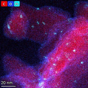
Materials have fundamentally different properties at the nanoscale than at the macroscale. To study them, S/TEM instrumentation can be combined with energy dispersive X-ray spectroscopy to obtain nanometer, or even sub-nanometer, resolution data.

Micro-traces of crime scene evidence can be analyzed and compared using electron microscopy as part of a forensic investigation. Compatible samples include glass and paint fragments, tool marks, drugs, explosives, and GSR (gunshot residue).

Catalysts are critical for a majority of modern industrial processes. Their efficiency depends on the microscopic composition and morphology of the catalytic particles; EM with EDS is ideally suited for studying these properties.

The diameter, morphology and density of synthetic fibers are key parameters that determine the lifetime and functionality of a filter. Scanning electron microscopy (SEM) is the ideal technique for quickly and easily investigating these features.

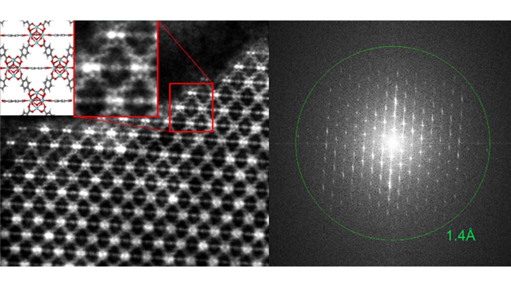
Novel materials research is increasingly interested in the structure of low-dimensional materials. Scanning transmission electron microscopy with probe correction and monochromation allows for high-resolution two-dimensional materials imaging.

Every component in a modern vehicle is designed for safety, efficiency, and performance. Detailed characterization of automotive materials with electron microscopy and spectroscopy informs critical process decisions, product improvements, and new materials.
For other products from ThermoFisher, click here.
