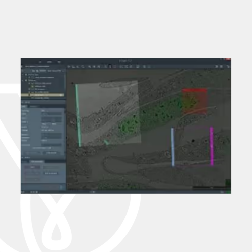
PT Wadya Prima Mulia as the Authorized Distributor for ThermoFisher Scientific in Indonesia, provides MAPS 3 Software for Electron Microscopes
Correlative electron microscopy and cross-platform imaging automation software.
For more information regarding the product, click here
Correlative Electron Microscopy Software
Thermo Scientific Maps Software is an imaging and correlative workflow software suite compatible with the full line of Thermo Scientific SEM, DualBeam (FIB SEM) and TEM platforms.
Scientists and researchers rely increasingly on nanoscale observations to inform the latest advances in research and analysis. It has, however, become apparent that high-resolution observations lose much of their utility in the absence of the larger macroscopic context. Observations from multiple sources must be linked, providing the necessary multi-scale and multi-modal insight for truly valuable data.
Maps Software provides a powerful imaging workflow automation package within an easy-to-use and robust platform. With just a few clicks, you can collect impactful data while preserving the context of your observations.
Multi-scale imaging automation
Whether its scanning electron microscopy analysis in SEM and DualBeam tools or transmission electron microscopy (TEM), Maps Software enables automated 2D imaging for a variety of applications, from acquiring large image mosaics to scheduling routine imaging tasks for off times (overnight, weekends).
- Acquire up to four simultaneous signals, automatically (STEM/SEM)
- Run multiple samples in series to increase system productivity
- Choose rectangular, circular or custom shapes for mapping acquisition (STEM/SEM)

Correlative microscopy
With the ability to correlate optical and electron microscope data and easy navigation from SEM to TEM, Maps Software allows you to obtain the necessary context that drives your research and development. Use multi-modal data to aid in interpretation and navigation, ensuring you are always collecting the right data from the right location.
- Import 2D and 3D data from any source
- Quickly generate context for the entire sample grid or targeted region of interest
- Register imagery from other modalities (e.g. energy-dispersive spectroscopy, secondary electron signal, electron backscatter diffraction, etc.)
- Integrate observations in a multi-scale, multi-layered visualization environment
- Align data with system stages using any Thermo Scientific EM platform

Integrated analytics
Maps Software supports integrated EDS map acquisition on STEM platforms for chemical characterization and elemental analysis along with your high-resolution imaging.
- Automate large-area, high-throughput EDS acquisition with the Thermo Scientific Super-X and Dual-X Detector Systems
- Enable native correlation with electron imaging layers
- Employ built-in stitching and multiscale visualization
- Easily view EDS maps away from the microscope with the optional offline version of Maps Software
Intuitive visualization and offline collaboration
The optional offline version of Maps Software lets you take the microscope with you when you are away from the lab.
- View your data from your own PC
- Access the full correlative power of Maps Software anywhere
- Plan for your next imaging session before you head to the lab
- Easily share your multi-scale observations with colleagues
Avizo Software integration
The addition of Thermo Scientific Avizo Software automates your image processing and statistical data generation. Avizo Software provides a rich, integrated user experience that blends acquisition with advanced analysis, and works in the background to process images as they are acquired.
- Gain instant access to critical statistical data through a complete, automated imaging and analysis solution
- Perform advanced image processing, accessible while you are on the tool
- Tune and optimize imaging and processing workflows

- Compatibility with current Thermo Scientific SEM, FIB-SEM, and TEM platforms; providing a unified user experience across all instrument types.
- Integrated approach to data acquisition, annotation and storage.
- Automated acquisition of large overviews at any magnification (tile and stitch); multiple acquisitions can be set up for unsupervised batch data collection.
- Setup and coordination of experiments across multiple microscopes.
- Correlation of images from different microscopes, including imported images from 3rd party instruments using the integrated Bio-Formats Library.
Multi-scale imaging automation
A completely automated solution for generating high-quality, multi-scale images.
Correlative microscopy
Gain insight by integrating observations across modalities and imaging platforms.
Integrated analytics
Make EDS mapping easier and instantly valuable via direct acquisition and integration of elemental layers.
Intuitive visualization and offline collaboration
The optional offline version of Maps Software allows you to continue your investigation and plan your next imaging session while away from the microscope.
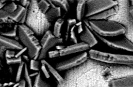
Process control using electron microscopy
Modern industry demands high throughput with superior quality, a balance that is maintained through robust process control. SEM and TEM tools with dedicated automation software provide rapid, multi-scale information for process monitoring and improvement.
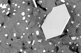
Quality control and failure analysis
Quality control and assurance are essential in modern industry. We offer a range of EM and spectroscopy tools for multi-scale and multi-modal analysis of defects, allowing you to make reliable and informed decisions for process control and improvement.
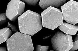
Fundamental Materials Research
Novel materials are investigated at increasingly smaller scales for maximum control of their physical and chemical properties. Electron microscopy provides researchers with key insight into a wide variety of material characteristics at the micro- to nano-scale.
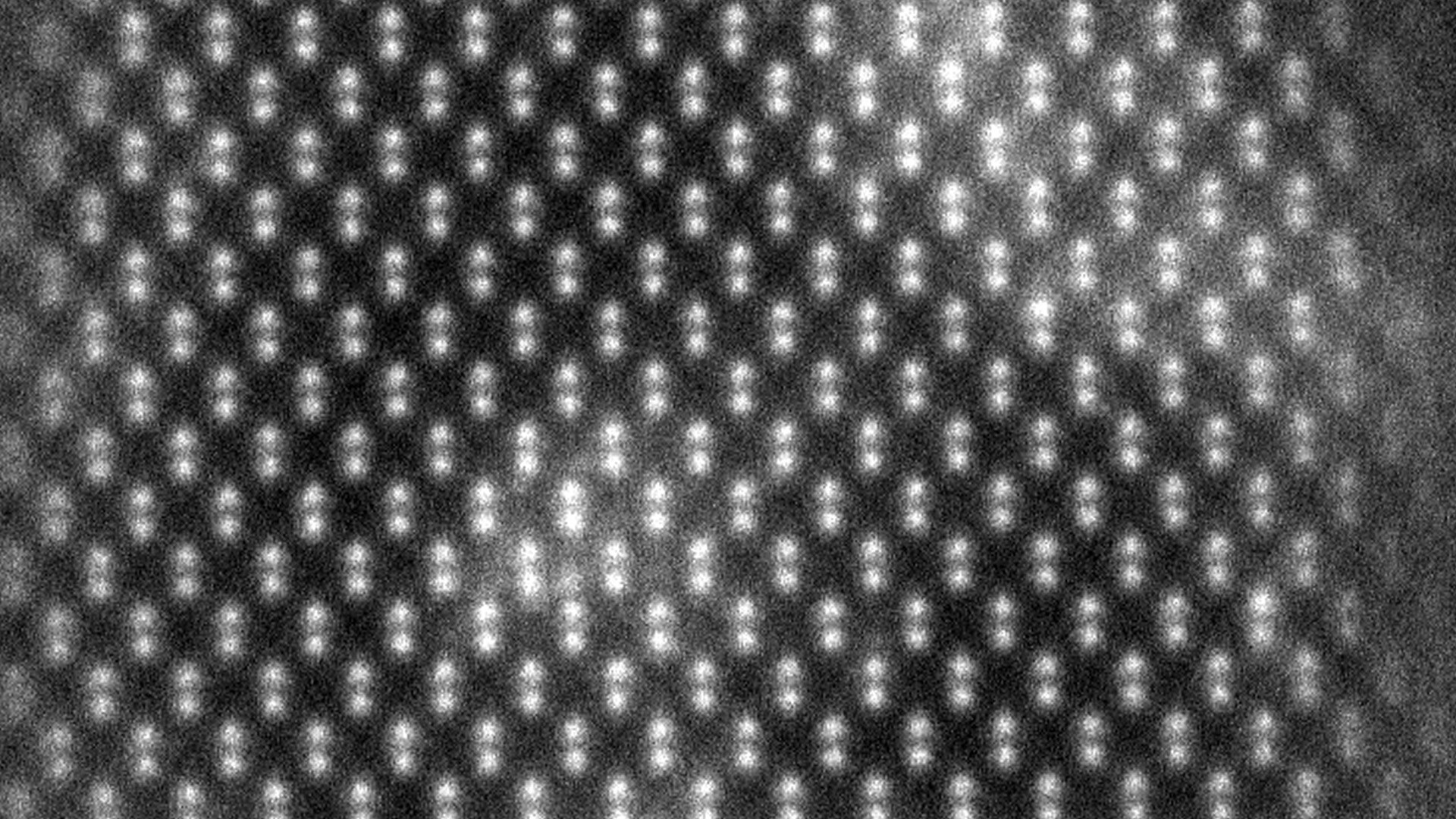
Semiconductor research and development
Innovation starts with research and development. Learn more about solutions to help you understand innovative structures and materials at the atomic level.
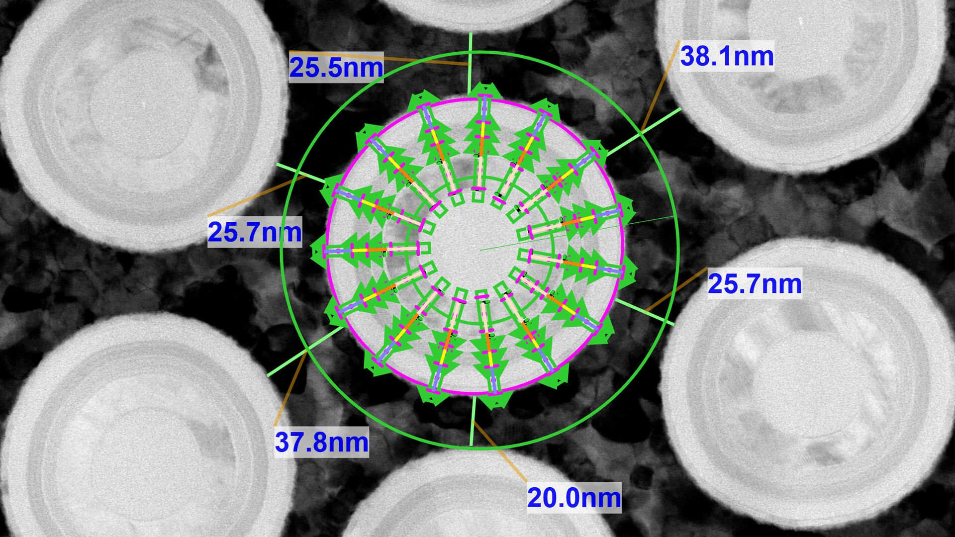
Manufacturing today’s complex semiconductors requires exact process controls. Learn more about advanced metrology and analysis solutions to accelerate yield learnings.
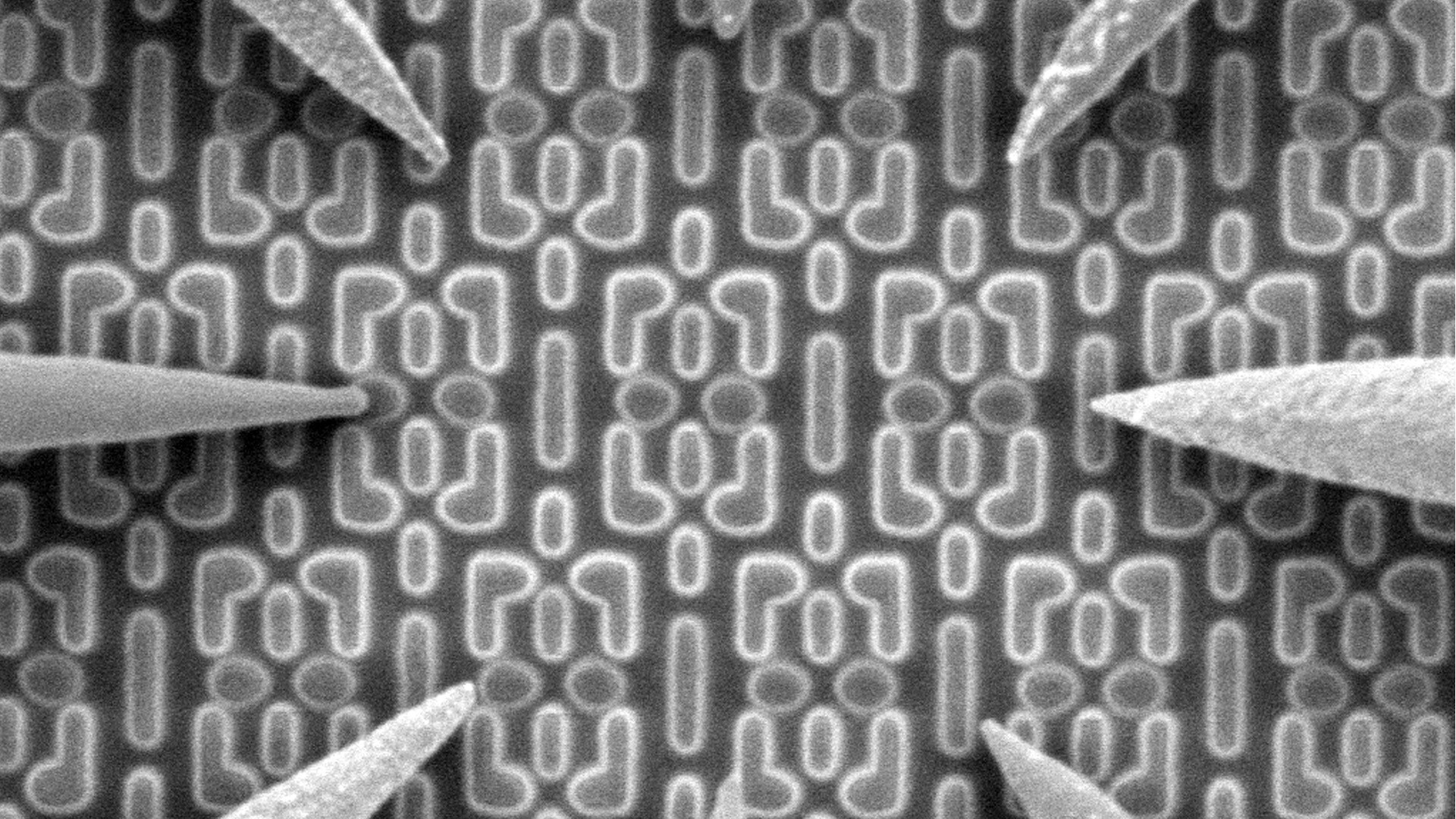
Semiconductor Failure Analysis
Complex semiconductor device structures result in more places for defects to hide. Learn more about failure analysis solutions to isolate, analyze, and repair defects.
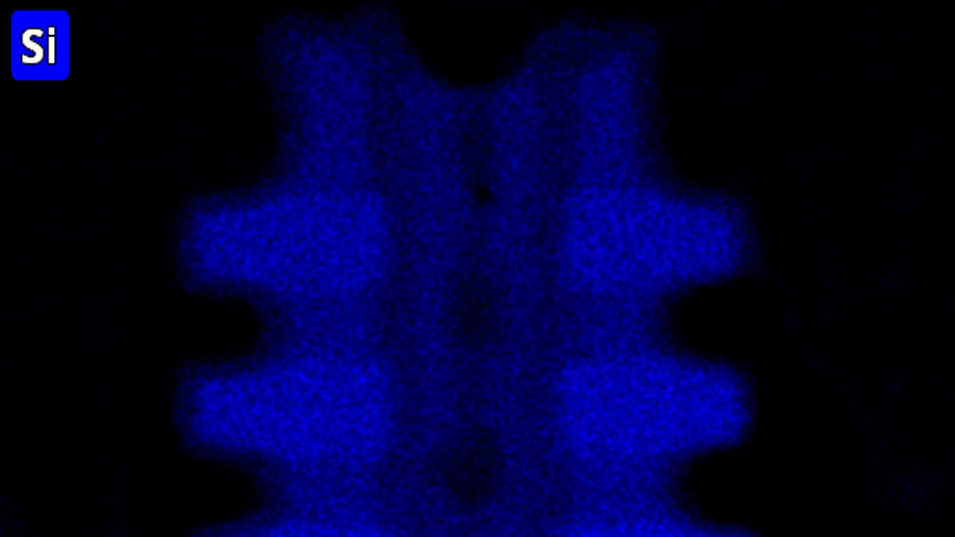
Semiconductor materials characterization
Many factors impact yield, performance, and reliability. Learn more about solutions to characterize physical, structural, and chemical properties.

Cryo-EM techniques enable multiscale observations of 3D biological structures in their near-native states, informing faster, more efficient development of therapeutics.
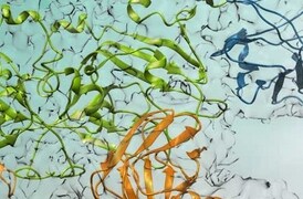
Learn how to take advantage of rational drug design for many major drug target classes, leading to best-in-class drugs.
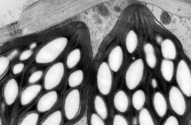
Fundamental plant biology research is enabled by cryo electron microscopy, which provides information on proteins (with single particle analysis), to their cellular context (with tomography), all the way up to the overall structure of the plant (large volume analysis).
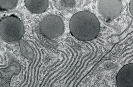
Transmission electron microscopy (TEM) is used when the nature of the disease cannot be established via alternative methods. With nano-biological imaging, TEM provides accurate and reliable insight for certain pathologies.
For other products from ThermoFisher, click here.
