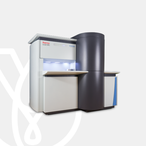
K-Alpha X-ray Photoelectron Spectrometer
The Thermo Scientific Scios 2 DualBeam is an ultra-high-resolution analytical focused ion beam scanning electron microscopy (FIB-SEM) system that provides outstanding sample preparation and 3D characterization performance for a wide range of samples, including magnetic and non-conductive materials. With innovative features designed to increase throughput, precision, and ease of use, the Scios 2 DualBeam is an ideal solution to meet the needs of scientists and engineers in advanced research and analysis across academic, governmental, and industrial research environments.
Subsurface characterization
Subsurface or three-dimensional characterization is often required to better understand the structure and properties of a sample. The Scios 2 DualBeam, with optional Thermo Scientific Auto Slice & View 4 (AS&V4) Software, allows for high-quality, fully automated acquisition of multi-modal 3D datasets, including backscattered electron (BSE) imaging for maximum materials contrast, energy-dispersive spectroscopy (EDS) for compositional information, and electron backscatter diffraction (EBSD) for microstructural and crystallographic information. Combined with Thermo Scientific Avizo Software, the Scios 2 DualBeam delivers a unique workflow solution for high-resolution, advanced 3D characterization and analysis at the nanometer scale.
Backscattered electron and secondary electron imaging
The innovative NICol electron column provides the foundation of the system’s high-resolution imaging and detection capabilities. It offers excellent nanoscale details, with a wide range of working conditions, whether operating at 30 keV in STEM mode (to access structural information) or at lower energies (to obtain charge-free, detailed surface information). With its unique in-lens Thermo Scientific Trinity Detection System, the Scios 2 DualBeam is designed for simultaneous acquisition of angular and energy-selective secondary electron (SE) and BSE imaging. Fast access to detailed nanoscale information is possible not only top-down, but also on tilted specimens or cross-sections. Optional below-the-lens detectors and an electron-beam-deceleration mode ensure fast and easy simultaneous collection of all signals, revealing the smallest features in a material surface or cross-section. Fast, accurate, and reproducible results are obtained thanks to the unique NICol column design with full auto alignments.
TEM sample preparation
Scientists and engineers constantly face new challenges that require highly localized characterization of increasingly complex samples with ever smaller features. The latest technological innovations of the Scios 2 DualBeam, in combination with the optional, easy-to-use, comprehensive Thermo Scientific AutoTEM 4 Software and Thermo Fisher Scientific’s application expertise, allow for fast and easy preparation of site-specific high-resolution S/TEM samples for a wide range of materials. In order to achieve high-quality results, final polishing with low-energy ions is required to minimize surface damage on the sample. The Thermo Scientific Sidewinder HT Focused Ion Beam (FIB) column not only delivers high-resolution imaging and milling at high voltages but also offers good low-voltage performance, enabling the creation of high-quality TEM lamella.
High-performance X-ray source
The X-ray monochromator allows selection of analysis area from 50 µm to 400 µm in 5 µm steps, fitting it to the feature of interest to maximize the signal.

Optimized electron optics
The high-efficiency electron lens, hemispherical analyzer, and detector allow for superb detectability and rapid data acquisition.

Sample viewing
Bring sample features into focus with the K-Alpha XPS System’s patented optical viewing system and XPS SnapMap, which helps you pinpoint areas of interest quickly.

Insulator analysis
The patented dual-beam flood source couples low-energy ion beams with very low energy electrons (less than 1 eV) to prevent sample charging during analysis, which eliminates the need, in most cases, for charge referencing.

Depth profiling
Go beyond the surface with the EX06 ion source. Automated source optimization and gas handling ensure excellent performance and experimental reproducibility.

Digital Control
Intuitive operation—guided by the Avantage data system—makes the K-Alpha XPS System ideal for both multi-user, shared facilities and XPS experts who place a premium on efficient operation and high-throughput analysis.

Optional sample holders
Specialist sample holders for angle-resolved XPS, sample bias measurements, or for inert transfer from a glove box are available.

| Analyzer type | 180° double-focusing hemispherical analyzer, 128-channel detector |
| X-ray source type | Monochromated, micro-focused, low-power Al K-Alpha X-ray source |
| X-ray spot size | 50–400 µm (adjustable in 5 µm steps) |
| Depth profiling | EX06 ion source |
| Maximum Sample area | 60×60 mm |
| Maximum sample thickness | 20 mm |
| Vacuum system | Two turbo molecular pumps, with automated titanium sublimation pump and backing pump |
| Optional accessories | ADXPS sample holder, work function sample holder, glove box |
Battery development is enabled by multi-scale analysis with microCT, SEM and TEM, Raman spectroscopy, XPS, and digital 3D visualization and analysis. Learn how this approach provides the structural and chemical information needed to build better batteries.

Quality control and assurance are essential in modern industry. We offer a range of EM and spectroscopy tools for multi-scale and multi-modal analysis of defects, allowing you to make reliable and informed decisions for process control and improvement.

Polymer microstructure dictates the material’s bulk characteristics and performance. Electron microscopy enables comprehensive microscale analysis of polymer morphology and composition for R&D and quality control applications.

Geoscience relies on consistent and accurate multi-scale observation of features within rock samples. SEM-EDS, combined with automation software, enables direct, large-scale analysis of texture and mineral composition for petrology and mineralogy research.

As the demand for oil and gas continues, there is an ongoing need for efficient and effective extraction of hydrocarbons. Thermo Fisher Scientific offers a range of microscopy and spectroscopy solutions for a variety of petroleum science applications.

Materials have fundamentally different properties at the nanoscale than at the macroscale. To study them, S/TEM instrumentation can be combined with energy dispersive X-ray spectroscopy to obtain nanometer, or even sub-nanometer, resolution data.

Micro-traces of crime scene evidence can be analyzed and compared using electron microscopy as part of a forensic investigation. Compatible samples include glass and paint fragments, tool marks, drugs, explosives, and GSR (gunshot residue).
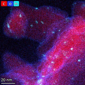
Catalysts are critical for a majority of modern industrial processes. Their efficiency depends on the microscopic composition and morphology of the catalytic particles; EM with EDS is ideally suited for studying these properties.

The diameter, morphology and density of synthetic fibers are key parameters that determine the lifetime and functionality of a filter. Scanning electron microscopy (SEM) is the ideal technique for quickly and easily investigating these features.
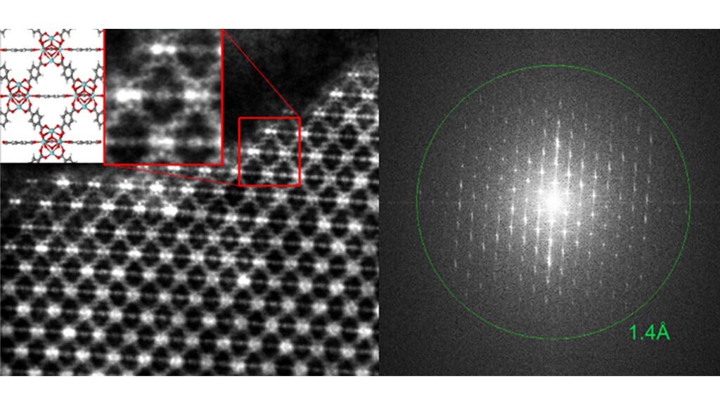
Novel materials research is increasingly interested in the structure of low-dimensional materials. Scanning transmission electron microscopy with probe correction and monochromation allows for high-resolution two-dimensional materials imaging.

Every component in a modern vehicle is designed for safety, efficiency, and performance. Detailed characterization of automotive materials with electron microscopy and spectroscopy informs critical process decisions, product improvements, and new materials.
For other products from ThermoFisher, click here.