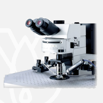
Olympus Upright Microscope BX61WI / BX51WI
PT Wadya Prima Mulia as the Exclusive Distributor for Evident-Olympus in Indonesia, provides BX61WI / BX51WI Upright Microscope and other products.
The BX51WI is ideal for all physiological experiments such as patch clamping and intravital microscopy. The fixed stage concept and vibration-free frame design ensure excellent stability throughout the experiment. The use of infrared light protects living cells and offers high penetration depths of thick tissue slices, while high NA optics allow magnification changes without moving the objective.
The BX61WI is the motorized version of the BX51WI fixed-stage microscope with a highly accurate Z-drive. It is the ideal tool for all automated physiological experiments, such as patch clamping and intravital microscopy.
Vibration-free Magnification Changes
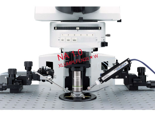
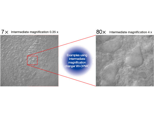
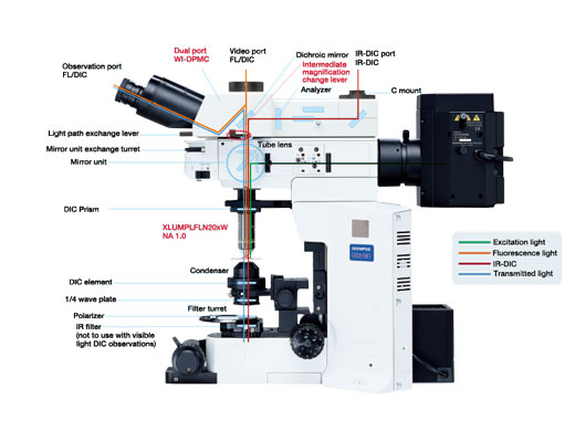
A major concern for researchers conducting electrophysiology experiments is the vibration which occurs when switching objectives and the resulting interference this can cause to the specimens and adjacent equipment. To solve this problem, Olympus introduces a concept – the provision of an intermediate magnification changer in combination with the High NA long working distance 20x objective that allows the user to switch between low and high magnifications without the need to switch objectives.
Safe Magnification Changes
The 20x water immersion objective (XLUMPLFLN20xW) makes high-resolution observation possible with a wide range of intermediate magnification lenses. Since exchanges between low and high magnification are performed through the intermediate magnification changer, vibration is reduced to a minimum and the usual concern about collisions between objectives and patch clamp electrodes is eliminated.
Simultaneous Fluorescence and IR-DIC Imaging
With the included 690 nm dichroic mirror in the WI-DPMC, fluorescence light is sent to the front port, and IR-DIC light is sent to the back port allowing two cameras to image simultaneously with no vibration introduced by light path selection. IR-DIC observation is compatible with 775 nm and 900 nm wavelengths.
Magnification Selector with Minimal Vibration
The WI-DPMC rear camera port includes a 2 position intermediate magnification selector. A high magnification 4x intermediate lens is included and a (0.25x or 0.35x) low magnification lens is optional. High or low magnification selection is via a single lever with no click-stops or detents allowing a specimen to be scanned and measured with minimal disturbance from vibration.
*Available for 0.5x, 1x and 2x intermediate magnification lenses by special order.
| Visible light DIC | Allows operator high-resolution observation of the tissue surface. |
| 775 nm IR-DIC | In combination with an IR camera observation within a tissue slice is made possible. Optics are corrected for visible and IR wavelengths allowing fast switching between wavelengths with minimal refocusing. |
| 900 nm Nomarski | Allows observation deeper into the tissue (requires a polarizer and analyzer optimized for 900 nm). |
Senarmont Compensation for Nomarski DIC Imaging
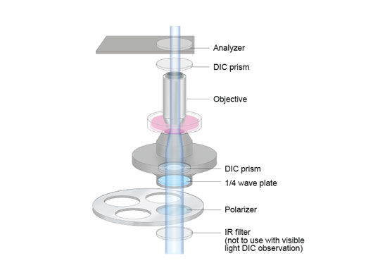
When using a Senarmont equipped condenser, all contrast adjustments are performed with the 1/4 wave plate below the condenser, thus eliminating the risk of bumping the stage, specimen, manipulators or nosepiece.
Variable Click-stops with Minimal Vibration
All click-stops, as when selecting between camera and observation modes, can be adjusted to have no click and thus no vibration.
Ultimate Clarity for Live Cell Electrophysiology
IR-DIC Optimized Optics
Thanks to precisely aberration-compensated IR-DIC optics, covering visible, 775 nm and 900nm wavelength near infrared light, the clarity of images observed under near infrared light has been further improved, allowing clear observation of deep sections of brain slices.
Condenser with DIC for Improved Contrast
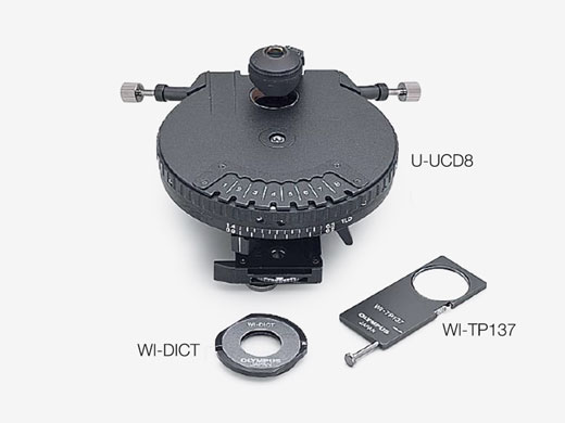
Suitable for use in visible and 775 nm to 900 nm near-infrared light, the U-UCD8 universal condenser is a high NA, short working distance condenser offering improved contrast in nerve cell observations, for example. The WI-UCD and WI-DICD offer solutions to various samples which needs long working distance.
Oblique Illumination for Optimized Contrast
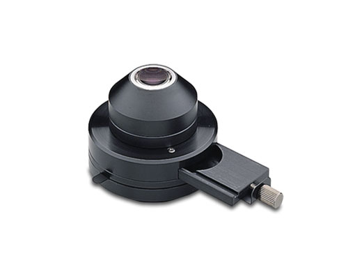
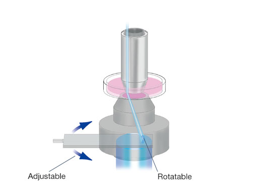
Olympus has developed an oblique condenser (WI-OBCD) whose long working distance enables the angles of shadow to be altered through 360 degrees without moving the specimen. Requiring no additional accessories, oblique illumination is easy to set up and control. Plastic dishes (normally unsuitable for all types of DIC) are easy to image with oblique illumination. The oblique illumination slit aperture is variable in size and on a slider allowing quick changeover.
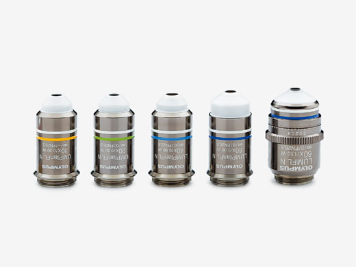
Objectives for long working distance
Dipping objectives designed with long working distances and special angles. Developed for experiments in electrophysiology.
Macro Lenses and Mirror Unit for Fluorescence
2x and 4x low magnification fluorescence objectives and a special GFP observation mirror unit are available. The objectives have a long working distance for maximum flexibility. An optional water immersion cap (XL-CAP) is also available to remove image aberrations caused by ripples on the water surface of immersed specimens.
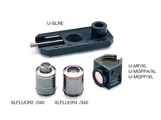
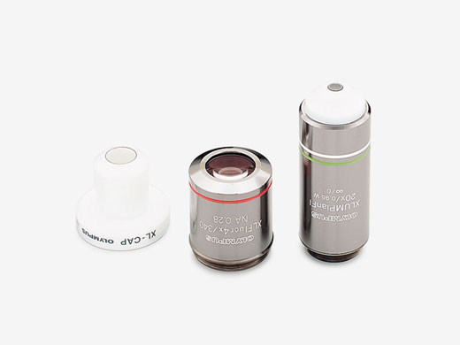
Objectives for Measuring Membrane Potential
The XLUMPLFLN20×W objective, with its high NA, and 2.0 mm of working distance allows the measurement of cell membrane electric potential (as seen left). Also, the 4x macro objective (XLFLUOR4×/340) can be used to measure membrane potential at the tissue level. A water immersion cap (XL-CAP) can be attached to the macro 2x or 4x objectives to eliminate disturbances caused by water ripples.
Vibration-free Minimal Noise Operation
The front operation system prevents interference in patch clamping work. The design concept is simple and allows frequently performed operations such as focusing or filter exchange to be quickly done at the front of the unit. Ample space is provided on both sides of the microscope frame and condenser, so the necessary manipulation equipment can be positioned close to the microscope.
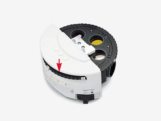
Vibration-free Shutter
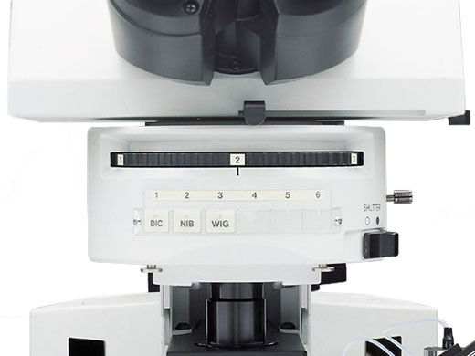
The fluorescence shutter slides horizontally with no detents or vibration.
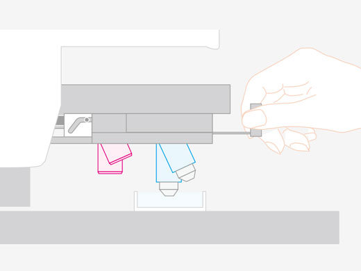
Mirror Unit Turret with Adjustable Click Release
The click-stop on the 6 position turret can be released with a screwdriver.
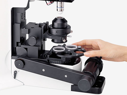
Designed for Easy Adjustments Around the Condenser
Frame designed for ample space around the condenser, making it easy to adjust Nomarski DIC contrast, exchange filters, adjust the condenser’s aperture stop and to easily switch between visible light, Nomarski DIC and IR-DIC.
Focus Knobs Close to Hand
Fine focus control is located at the front on both sides of the microscope body
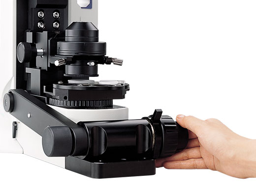
Lockable Coarse Focus
When engaged at the desired position, the objective can be raised with the coarse focus knob and then returned precisely to its original position.
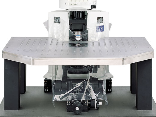
A Waterproofing Sheet to Protect Components
A waterproofing sheet, attached by the supplied magnets, provides protection against liquid overflow and spills. The sheet is large enough to protect the frame, condenser and focusing mechanisms.
Remote Power Supply and Hand Switch
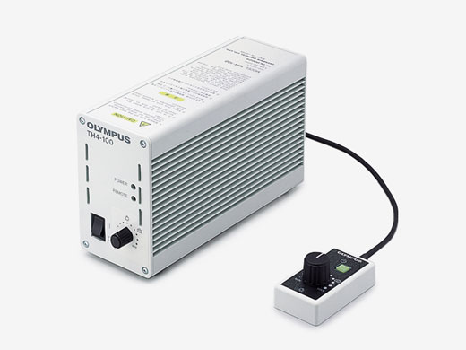
The remote TH4 power supply for transmitted light is designed with no cooling fan to minimize electrical noise. Features on/off and intensity controls. Can also be used with the optional TH4-HS hand switch providing light intensity and on/off control a maximal distance away from the Faraday cage.
A Wide Choice of Nosepieces
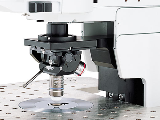
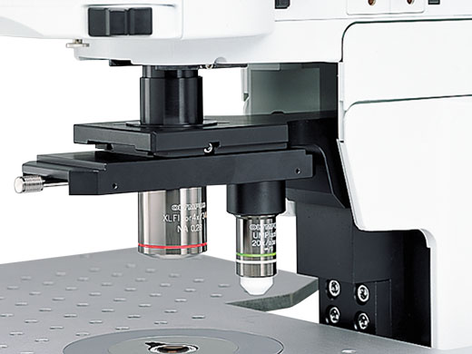
The Swing nosepiece WI-SRE3 has a unique slim, compact design and front-to-back swing motion to enable objective changes without interfering with electrodes and micromanipulators. Objective positioning incorporates a vibration-free counter spring mechanism. The Slide nosepiece U-SLRE is designed for the attachment of one large diameter, low magnification fluorescence objective (XLFLUOR 2x/340 or 4x/340) and one objective with normal (RMS) diameter threads. Nosepiece motion is a simple horizontal slide. The Single position nosepiece WI-SNPXLU2 is designed to accept the unique, large diameter XLUMPLFLN20×W objective. The RMS adapter WI-RMSAD enables the attachment of an objective with RMS thread size to the WI-SNPXLU2.
The Swing-slide Nosepiece Avoids Air Bubbles
This nosepiece features a swing-slide motion, whereby the objective swings forward while being raised. As a result, the objective clears the walls of the perfusion chamber. This motion also prevents the trapping of air bubbles when the objective is lowered.
Left and Right Handed Operation
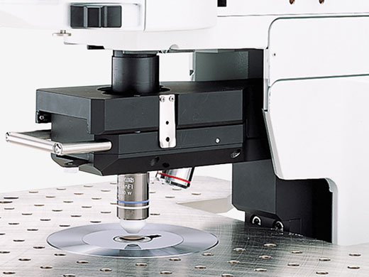
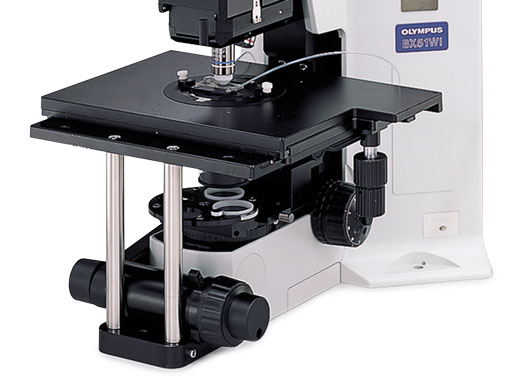
The IX-SVL2 general purpose platform stage mounts for left or right handed operation and enables stable specimen X-Y movement.
Functionality and Solutions for a Range of Needs
Adjustable for Small Animal Experiments
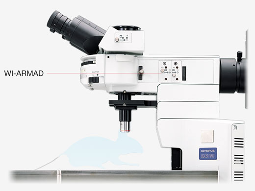
The arm height raising kit (WI-ARMAD) provides an additional 40 mm of clearance and is mounted between the microscope frame and the reflected light illuminator. Small animal experiments usually do not require transmitted light thus allowing the removal of the substage condenser assembly. After removal, the stage may be lowered an additional 50 mm, providing a total clearance increase of 90 mm.
Additional Units to Add and Control Light Sources
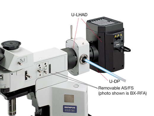
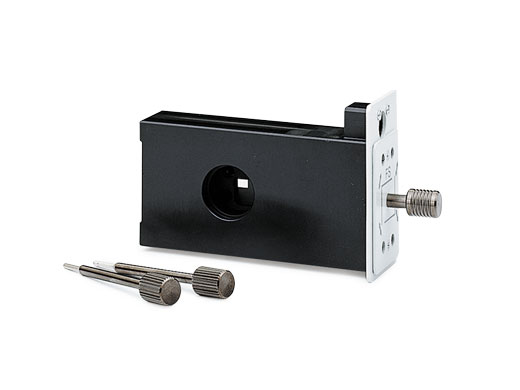
The Rectangular field stop U-RFSS is designed for use with CCD cameras and prevents photobleaching of the specimen outside of the imaging area.
BX Stage and Adapter for Injection Experiments
The stage adapter WI-STAD is designed to allow the attachment of a traditional microscope right or left hand stage to the WI frame. The compact design of the BX2 stage (U-SVRB-4, or U-SVLB-4) reduces the distance between the specimen and the manipulator and creates a stable platform for injections.
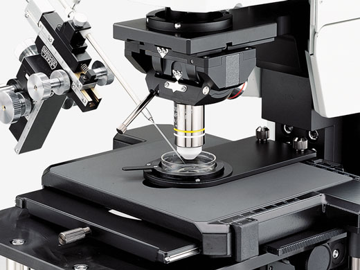
Accessories
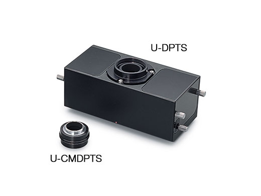
Double Port Tube to Split Visible and Infrared
The dual port tube U-DPTS accepts an optional dichroic mirror allowing the incoming light to be split between visible and infrared and be observed simultaneously using two cameras.
* A fluorescence mirror unit is required.
Intermediate magnification changer for IR
The U-ECA, which includes a 2x intermediate magnification position, allows quick magnification changes to a camera or observer without the need to change objectives. The U-CA includes a 4 position turret that allows rapid switching between a 1x, 1.25x, 1.6x and 2x positions. Both changers accept standard Olympus adapters for attaching a wide range of cameras.
* U-ECA and U-CA are not recommended for IR observation with the U-TR30 trinocular observation head.
C-mount Video Magnification Changer for IR
The U-TVCAC includes a 3-position turret with 1x, 2x, and 4x IR corrected positions. Includes a standard c-mount top port.
| Observation Method | Brightfield | ||
|---|---|---|---|
| Darkfield | |||
| Fluorescence (Blue/Green Excitations) | |||
| Fluorescence (Ultraviolet Excitations) | |||
| Differential Interference Contrast | |||
| IR-Differential Interference Contrast | |||
| Simple Polarized Light | |||
| Illuminator | Fluorescence Illuminator | Xenon Lamp |
|
| Focus | Focusing Mechanism | Nosepiece Focus | |
| Motorized |
|
||
| Intermediate Magnification Changer | Manual Terret | ||
| Stage | Manual | Manual Stages with Right-Hand Control |
|
|
Stage
|
Mechanical | IX-SVL2 Cross Stage with Short Left Handle |
|
| Condenser | Manual | Universal Condenser | NA 0.9/ W.D. 1.5 mm for 1.25X–100X [swing-out: 1.25X–4X, with oil top lens: (NA 1.4/ W.D. 0.63 mm)] |
| Swing-Out Condenser | NA 0.9/ W.D. 2 mm (1.25X–100X) | ||
| Long Working Distance Universal Condenser | NA 0.8/ W.D. 5.7 mm (10X–100X) | ||
| Long Working Distance DIC Condenser | NA 0.8/ W.D. 5.7 mm (10X–100X) | ||
| Long Working Distance Oblique Condenser | NA 0.8/ W.D. 5.7 mm (10X–100X) | ||
| Observation Tubes | Widefield (FN 22) | Trinocular | |
| Trinocular for Infrared | |||
| Erected Trinocular | |||
| Dimensions (W × D × H) | 317.5 (W) x 567 (D) x 503.8 (H) mm (Epifluorescence Configuration) | ||
| Weight | BX61WI: 21 kg (Epifluorescence configuration), BX51WI: 19 kg (Epifluorescence configuration) | ||
For other products from Evident-Olympus, click here.
